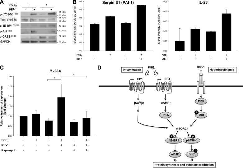
Fig. 7.
Potentiating cross talk between PGE2 and IGF-1 signaling pathways through mTORC1. A: PANC-1 cells were stimulated with IGF-1 (10 ng/ml) in the absence or presence of PGE2 (1 μM) for 15 min. After treatments, cell lysates were collected and subjected to immunoblotting with indicated antibodies. B: relative levels of plasminogen activator inhibitor (PAI)-1 and IL-23 in PANC-1 cell supernatants collected after 24 h of indicated treatments were determined using a cytokine array. Values, expressed as signal intensity (arbitrary units), are averages quantified from duplicated spots on the array membrane. C: after 24 h of treatment with PGE2, IGF-1, and rapamycin, relative transcript expression levels of IL-23 in PANC-1 cells were determined by RT-qPCR. *P < 0.05 (by Student's t-test). D: schematic model showing cross talk between the PGE2 and IGF-1 pathways converging at mTORC1 implicated in obesity-associated tumor promotion. [Ca2+]i, intracellular Ca2+ concentration; PI3K, phosphoinositide 3-kinase; eIF4E, eukaryotic translation initiation factor 4E; 4E-BP1, eIF4E-binding protein; p70S6K, ribosomal protein S6 kinase; S6rp, S6 ribosomal protein.

