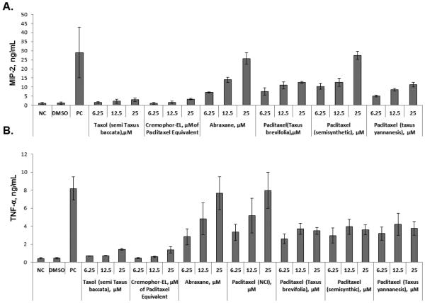
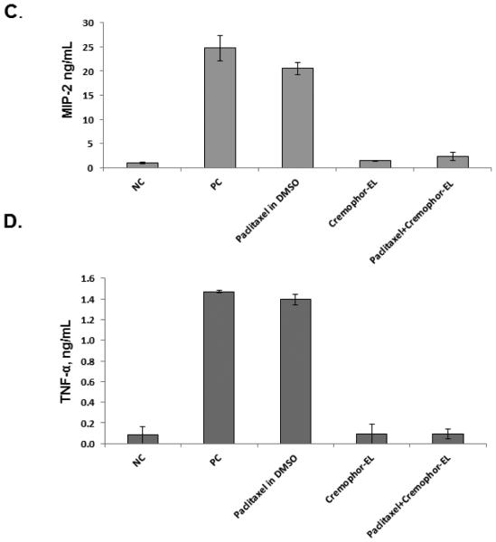
Figure 2. Effect of Cremophor-EL on paclitaxel-induced production of MIP-2 and TNF-α in murine macrophages.
Raw 264.7 cells were incubated with test samples and controls for 20 h, and the secretion of MIP-2 (A) and TNF-α (B) was analyzed by ELISA. PC – positive control (20 ng/mL of the LPS); NC – negative control (culture medium); DMSO (5.9 mg/mL) was used as a vehicle control for paclitaxels; test samples were Taxol®, Abraxane®, and paclitaxel from different sources dissolved in DMSO. The effects of Cremophor-EL on paclitaxel-triggered MIP-2 (C) and TNF-α (D) secretion by murine macrophages were evaluated by simultaneous addition of Cremophor-EL and 12.5 μM of paclitaxel in DMSO to the cells. Shown is the mean response and standard deviation from three independent experiments (N=3). Each sample within individual experiment was analyzed in duplicate (%CV <20).


