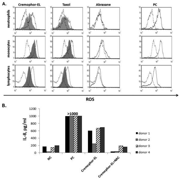
Figure 5. Cremophor-EL induces oxidative stress in human cells.
Human PBMCs were treated with the negative control (NC), the positive control (PC), Taxol®, Abraxane®, or Cremophor-EL with or without 5 mM of NAC. (A) After 1 h of treatment, cells were loaded with fluorescent dye sensitive to oxidative stress and analyzed by flow cytometry. The shift in green fluorescent channel FL-1 intensity (X-axis) is indicative of oxidative stress. Carbonyl cyanide m-chlorophenylhydrazone was used as the PC to induce oxidative stress. Shown is representative data from one of 4 tested donors (B) The levels of the IL-8 protein were tested in culture supernatants by ELISA 20 h after treatment of cells from the same donors used in A. The NC is cell culture media and the PC is 20 ng/mL of the LPS; Taxol® and Abraxane® were tested at equivalent (25 μM) concentrations of paclitaxel. The concentration of Cremophor-EL in Cremophor-EL-treated samples was equivalent to that in Taxol® when the Taxol® was used at 25 μM of paclitaxel. Each bar represents the mean value of the duplicate sample obtained from individual donor (N=2, %CV < 20%). Reference to the individual donor (#1 through #4) is provided in the box shown on the right.

