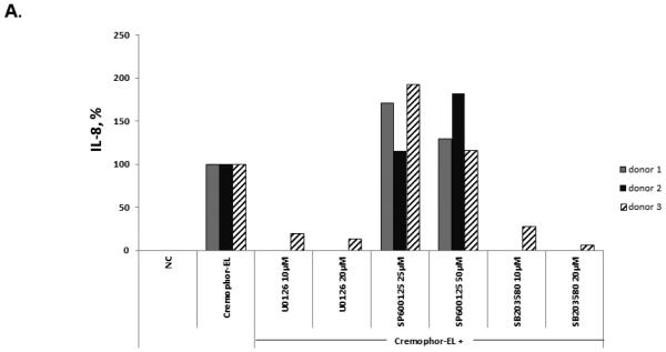
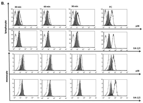
Figure 8. Effects of Cremophor-EL on MAPK.
(A) Human PBMCs were treated with 25 μM of Cremophor-EL and indicated concentrations of MAPK inhibitors. IL-8 was measured by ELISA in 20-h culture supernatants. Each bar represents the mean value of the duplicate sample obtained from individual donor (N=2, %CV < 20%). Reference to the individual donor (#1 through #3) is provided in the box shown on the right. Data are presented as the percentage of IL-8 protein induced by Cremophor-EL; the amounts of Cremophor-EL-triggered IL-8 were assigned to be 100%. (B) Human PBMCs were treated with 25 μM of Cremophor-EL for various time points before permeabilization and staining with antibodies specific to phosphorylated forms of p38 and Erk1/2. Dotted line – isotype control; filled histogram – NC; solid line – Cremophor-EL. Shown is the representative data from one of three individual donors.


