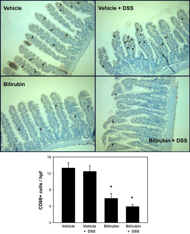
Fig. 10.
Monocyte infiltration into the small intestine is reduced following bilirubin treatment. Representative small intestinal specimens stained for CD68-positive cells (arrows) are shown (×200), as described under Fig. 8. Bottom: mean number of CD68-positive cells per hpf (±SE) for each treatment group. An average of 32 separate hpf was examined per specimen (n = 3 per group). *P < 0.01 vs. both vehicle treatment groups.

