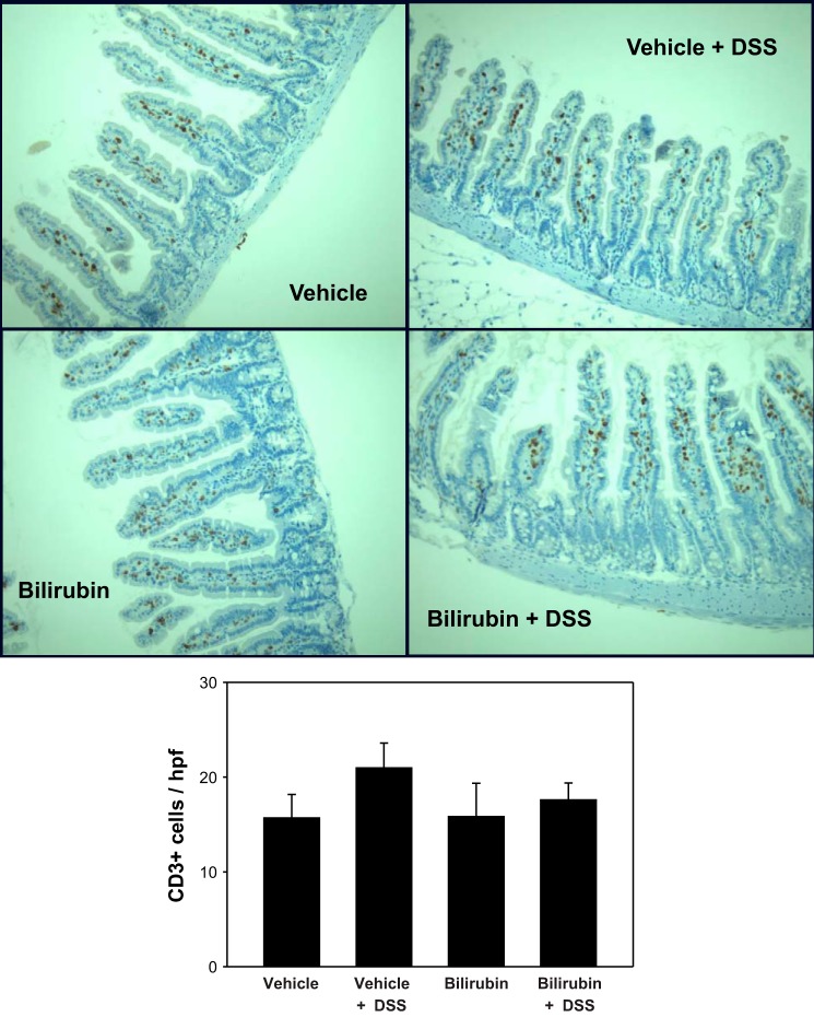
Fig. 9.
DSS and bilirubin do not alter T lymphocyte levels in the small intestine. Histological sections of small intestine (×200) were stained for CD3, as detailed in Fig. 7. Quantification of CD3-positive cells is represented as the number (±SE) per intact villus (bottom). An average of 32 villi was examined per specimen (n = 3 per treatment group).

