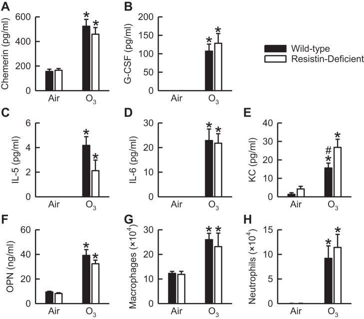
Fig. 4.
The concentration of chemerin (A), granulocyte-colony stimulating factor (G-CSF; B), IL-5 (C), IL-6 (D), keratinocyte chemoattractant (KC; E), and osteopontin (OPN; F) as well as the number of macrophages (G) and neutrophils (H) in bronchoalveolar lavage fluid (BALF) of wild-type C57BL/6 mice and mice genetically deficient in resistin (resistin-deficient mice) 24 h following cessation of a 3-h exposure to either filtered room air (air) or ozone (O3; 2 ppm). IL-5 and IL-6 were not detectable in BALF of wild-type and resistin-deficient mice exposed to air when using the immunoassay described in materials and methods. Each value is expressed as means ± SE; n = 6−8 mice in each group. *P < 0.05 compared with genotype-matched mice exposed to air; #P < 0.05 compared with resistin-deficient mice with an identical exposure.

