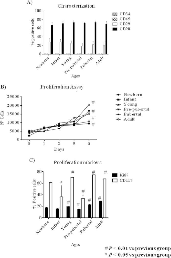
Figure 1. Proliferation profile from rat mesenchymal stem cells at different age.

(A) Characterization by flow cytometry assay of percentage of positives mesenchymal stem cells markers (CD29 and CD73) and negative hematopoietic markers (CD34 and CD45). (B) Proliferation assay of studied aging groups for 6 days. (C) Percentage of proliferation markers, DC117 and Ki67, from studied aging groups by flow cytometry assay. One representative experiment is shown. #p value less than 0.05 compared with previous group and *p value less than 0.01 compared with previous group, were considered statistically significant using Mann-Whitney-U tests.
