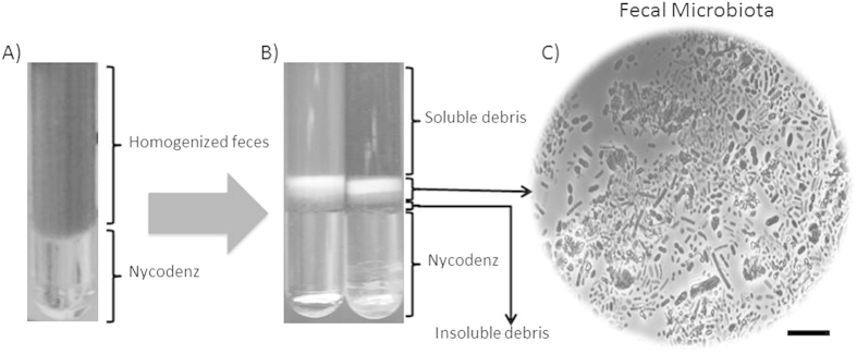
Figure 1. Workflow of the experimental setup used in this work.
(A) Diluted homogenized fecal samples were loaded on top of a Nycodenz® solution, as described in the material and methods section. (B) After centrifugation four layers were formed. Examination of the layer content in a phase-contrast microscope allowed us to determine the presence of one layer, corresponding to the fecal microbiota, between two layers containing soluble (upper) and insoluble (lower) fecal debris all above the Nycodenz. (C) Light photography of the microbiota layer, showing a high diversity of microbial sizes and shapes. Bar, 10 μm.

