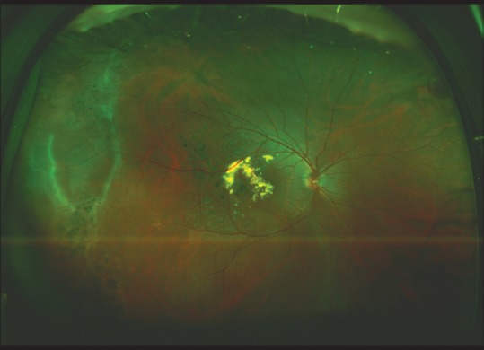
Figure 10.

An ultra-widefield color fundus photo of the right eye of a 13-year-old male with Coat's disease showing both peripheral and central retinal involvement. Laser photocoagulation was applied to ablate telangiectatic retinal vessels, which led to reduction in exudation
