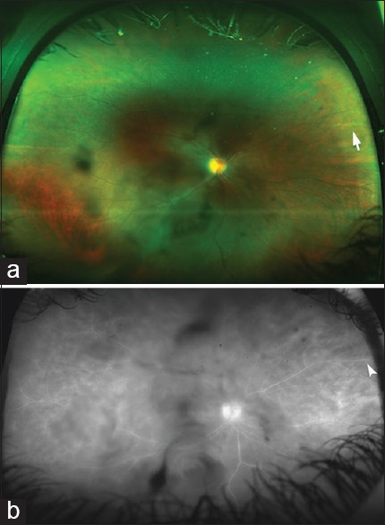
Figure 4.

(a) An ultra-widefield color fundus photo of the right eye of a 36-year-old patient with a history of pars planitis in both eyes. The image was taken with an ultra-wide imaging system (Optos 200Tx, Dunfermline, UK). Dark shadows in the central field represent vitreous opacities. There is a sclerotic retinal vessel in the nasal periphery (white arrow) in the area of prior inflammation, (b) an ultra-widefield fluorescein angiogram of the same eye as in a showing central vitreous opacities and staining of the peripheral retinal vessel (white arrowhead)
