Figure 5.

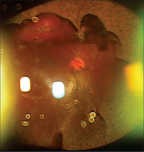
An ultra-widefield color fundus photo of an eye with proliferative diabetic retinopathy taken with slit-lamp and three-dimensional CCD camera. Inverted image shows preretinal hemorrhage inferiorly and scattered laser photocoagulation burns

An ultra-widefield color fundus photo of an eye with proliferative diabetic retinopathy taken with slit-lamp and three-dimensional CCD camera. Inverted image shows preretinal hemorrhage inferiorly and scattered laser photocoagulation burns