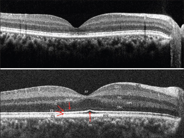
Figure 1.

An optical coherence tomography image from Case 1 (above) with unexplained vision loss shows disruption in the ellipsoid zone at the fovea with an intact external limiting membrane. An example of normal retina is represented below (CC: Choriocapillaries; EZ: Ellipsoid zone, FP: Foveal pit, FT: Foveal tent, GCL: Ganglion cell layer, INL: Inner nuclear layer, IPL: Inner plexiform layer, IS: Inner segment of the photoreceptor, NFL: Nerve fiber layer, OLM: Outer limiting membrane, ONL: Outer nuclear layer, OPL: Outer plexiform layer, OS: Outer segment of the photoreceptor, RPE: Retinal pigment epithelium complex)
