Figure 5.

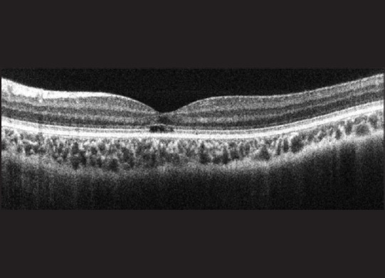
A spectral domain-optical coherence tomography image of Case 5 with complete achromatopsia showing a disrupted outer segment layer at the fovea demonstrating the hypo-reflective zone seen with cone dysfunction

A spectral domain-optical coherence tomography image of Case 5 with complete achromatopsia showing a disrupted outer segment layer at the fovea demonstrating the hypo-reflective zone seen with cone dysfunction