
Figure 9.
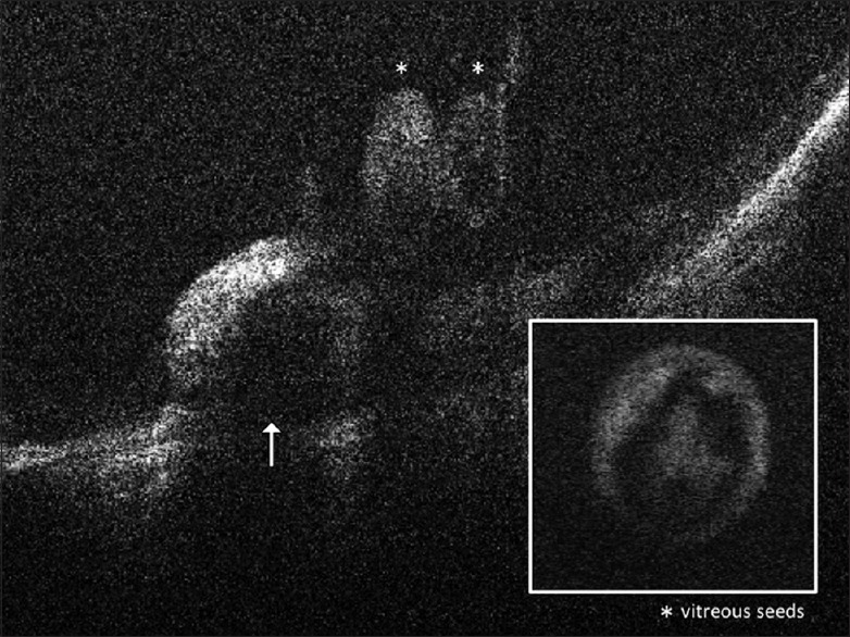
Spectral domain-optical coherence tomography images of a retinoblastoma tumor (indicated by an arrow) dispersing vitreous seeds (indicated by FNx01) into the vitreous. The box (inset) shows a single pearl-like vitreous seed with an outer layer of viable cells surrounding a core of necrotic cell debris