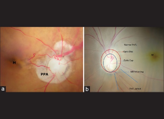
Figure 4.

(a) Optic nerve head of donor eye with glaucoma suspect. The image showing the right optic nerve head of the glaucoma suspect donor eye of a 96-year-old female with cup-disc ratio 0.9 (serial number 3 in Table 3). The parapapillary atrophy (PPA) suggestive of glaucoma is seen around the optic disc (×16). (b) Optic nerve head of donor eye with glaucoma suspect. The image showing the left optic nerve head of the glaucoma suspect donor eye of a 75-year-old male with cup-disc ratio 0.6 (serial number 9 in [Table 3]). The retinal rim thinning and retinal nerve fiber layer defect suggestive of glaucoma are seen in the stereoscopic fundus image (×16)
