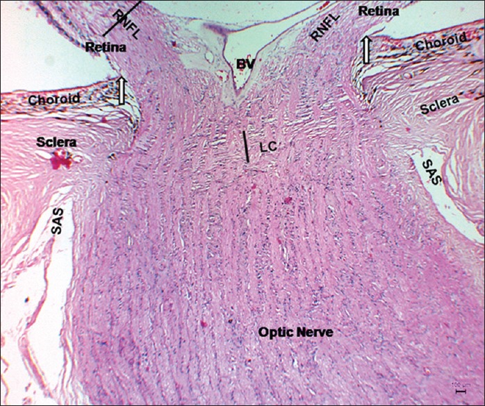
Figure 5.

Micrograph of sagittal section of optic nerve head of nonglaucomatous eye. The median sagittal section of a nonglaucomatous optic nerve head showing the absence of RNFL thinning, LC displacement, optic nerve head cupping and axonal loss. The vertical line represents the central thickness of LC. White arrow indicates the end of Bruch's membrane. Note: The RNFL and LC central thickness were 201.9 µm and 259.5 µm respectively. BV: Blood vessel; LC: Lamina cribrosa; SAS: Sub-arachnoids’ space, RNFL: Retinal nerve fiber layer (periodic Schiff-eosin)
