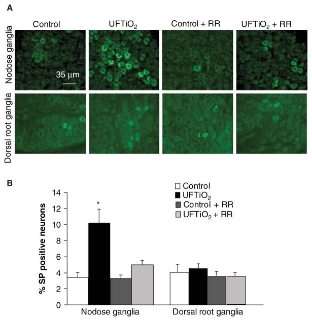
Figure 4.
Fluorescence photomicrographs of substance P (SP) immunoreactivity in nodose and dorsal root ganglia. (A) SP immunoreactivity was detected in different groups at 24 h post-exposure. (B) The SP immunoreactivity in nodose and dorsal root ganglia was quantified (B). Each value represents the mean ± SD of 5 rats; p < 0.05 compared with the control group (*).

