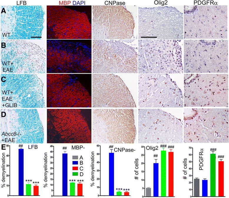
Fig. 6.

Glibenclamide and Abcc8−/− promote remyelination in EAE. a–d White matter of lumbar spinal cord sections from WT control (a), untreated pid-30 WT/EAE (b), glibenclamide-treated pid-30 WT/EAE (c), and pid-30 Abcc8−/−/EAE (d) mice, stained with Luxol fast blue (LFB) or immunolabeled for MBP (myelin), CNPase, Olig2 (oligodendrocytes), or PDGFR-α (oligodendrocyte progenitor cells (OPC)), as indicated; nuclei stained with DAPI in the MBP sections; original magnification, ×200 (LFB) or ×400 (all immunolabelings). e three left panels: Percent of quadrants with myelin loss by LFB staining, by MBP immunolabeling, or by CNPase immunolabeling. e two right panels: Quantification of Olig-2-expressing cells in white matter or of PDGFR-α-expressing OPC near the central canal; four mice/group; ## P < 0.01 and ### P < 0.001 with respect to WT control; ***P < 0.001 with respect to WT/EAE; scale bars, 100 μm
