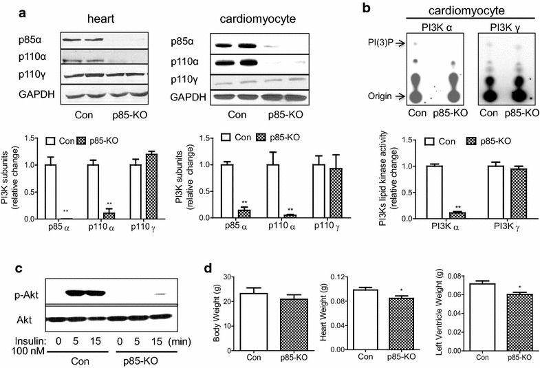
Fig. 1.

Characterization of p85-KO mouse hearts. a. Representative blots of p110α and p85 subunits of PI3Kα and p110 γ subunit of PI3Kγ in whole heart lysate (top left) and in freshly isolated adult mouse cardiomyocytes (top right). GAPDH was used as a loading control. Quantitative data of expression of PI3 K subunits in Con and p85-KO mouse hearts (n = 4) (bottom left) and cardiomyocytes (n = 7) (bottom right); b representative TLC plates show PI3Kα and PI3Kγ lipid kinase activities in freshly isolated Con or p85-KO cardiomyocytes (top). Quantitative data of PI3K activity in Con and p85-KO cardiomyocytes (n = 4); c insulin (100 nM)- induced Akt signaling in Con and p85-KO cardiomyocytes (representative blots of three experiments) and d comparison of body weight, heart weight and left ventricular weight in Con and p85-KO mice (Con n = 5, p85-KO n = 7). *P < 0.05, **P < 0.01 vs. Con
