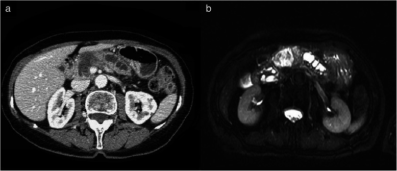
Fig. 1.

a Contrast enhanced CT scan demonstrates a complex pancreatic macrocystic mass involving the neck and the body of the pancreas with a peripheral solid portion. b Axial T2-weighted MRI shows the high-intensity central cystic portion of the mass with inner irregular septa and the peripheral less intense solid tissue determining main duct dilatation
