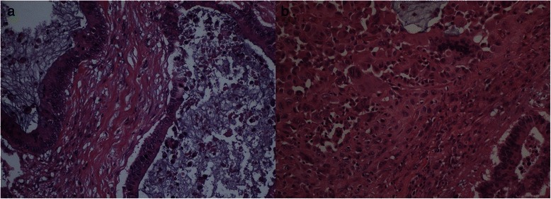
Fig. 2.

a Microscopically the tumor is characterized by a predominant cystic lesion composed of a columnar mucinous epithelium with atypical nuclei, numerous mitoses and stromal invasion. An ovarian-type stroma is absent (hematoxylin/eosin staining, 20x). b The solid area contains mononuclear spindle-shaped cells, pleomorphic giant cells and scattered multinucleated osteoclast-like giant cells (hematoxylin/eosin staining, 20x)
