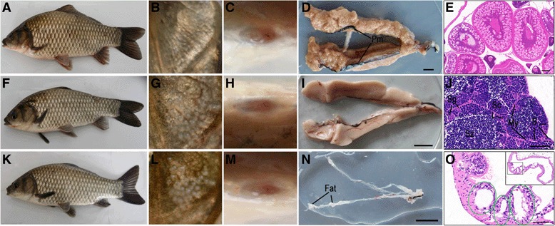
Fig. 5.

Phenotypic masculinization and their gonadal morphology in the germ cell-depleted adults. a-e, f-j and k-o Representative images of control gynogenetic females, control males from sexual reproduction, and the germ cell-depleted gynogenetic males, respectively. a, f and k Body shape. b, g and l Gill cover. c, h and m Anus. d, i and n Images of mature ovary, testis, and 1-year-old germ cell-depleted gonad, respectively. e, j, o HE staining of control ovary, control testis, and 1-year-old germ cell-depleted gonad, respectively. Green circle indicates the cavity, inset shows the whole cross section of 1-year-old germ cell-depleted gonad. Pm, Peritoneal membrane; Sg, spermatogonia; Sc, spermatocytes; Sz, spermatozoa; L, Leydig cells; S, Sertoli cells; My, peritubular myoid cells. [Scale bars, D, I and N, 2 cm; E 100, μm, J and O 50 μm]
