
Figure 1.
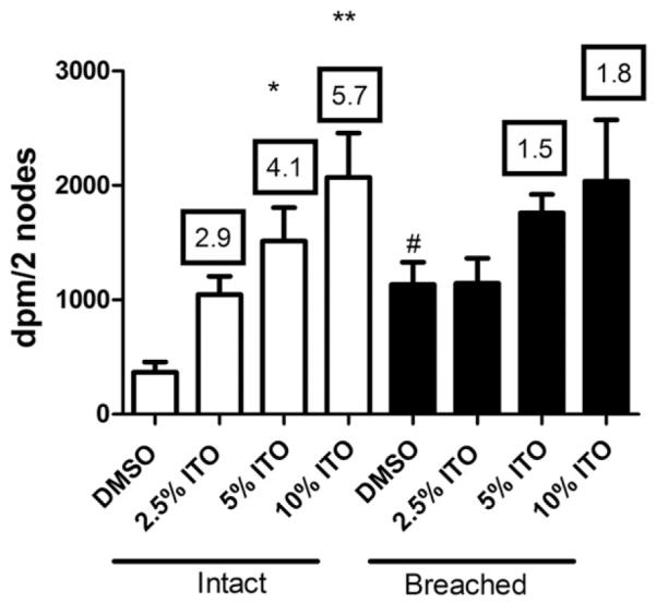
Lymphocyte proliferation. Proliferation was assessed by [3H]-thymidine incorporation into draining lymph node cells following dermal intact or breached skin exposure to vehicle (DMSO) or uITO. Bars represent means ± SE of five mice/group. Numbers appearing above the bars are the stimulation indices for respective concentration tested. Levels of statistical significance are denoted by *p<0.05 and **p<0.01 as compared to corresponding vehicle; #designates significant difference between animals exposed to DMSO or the same concentration of test article through breached vs intact skin. An EC3 of 4.7% was calculated for exposure to intact skin.