
Figure 2.
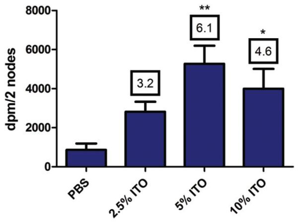
Lymphocyte proliferation. Proliferation was assessed by [3H]-thymidine incorporation into draining lymph node cells following intradermal exposure to vehicle (PBS) or uITO. Bars represent means ± SE of five mice/group. Numbers appearing above the bars are the stimulation indices for respective concentration tested. Levels of statistical significance are denoted by *p<0.05 and **p<0.01 as compared to vehicle control.