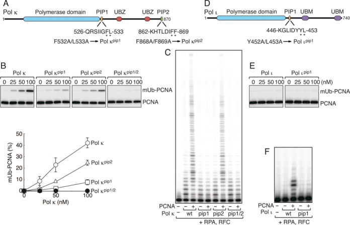
Figure 4.

Analysis of Polκ, Polι and their pip mutants in vitro. (A, D) Schematic structures of human Polκ (A) and Polι (D), as shown in Figure 1A. (B, E) PCNA ubiquitination assays of His-Polκ (B) and Polι (E), as shown in Figure 2. (C, F) DNA polymerase assays of His-Polκ (C) and Polι (F), as shown in Figure 3.
