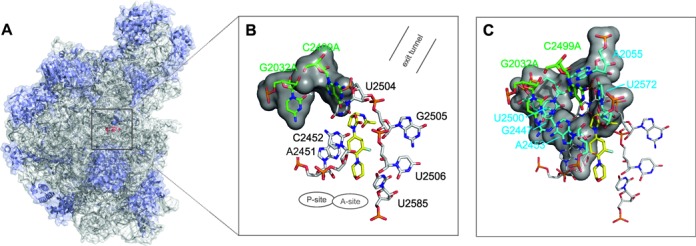
Figure 1.

Binding region of linezolid in H50S. (A) Structure of the large ribosomal subunit (PDB code 3CPW (8)). The ribosomal RNA is shown in gray and the protein chains are shown in blue; the binding position of linezolid (red) is depicted by a black square. (B and C) Binding mode of linezolid in the PTC of H50S. Nucleotides forming the first (black labels) and second shell (light blue labels) of the binding site are depicted in B and C, respectively; the two mutation sites (G2032A and C2499A) are highlighted in green. The locations of the A- and P-site and of the exit tunnel are indicated.
