FIGURE 5.

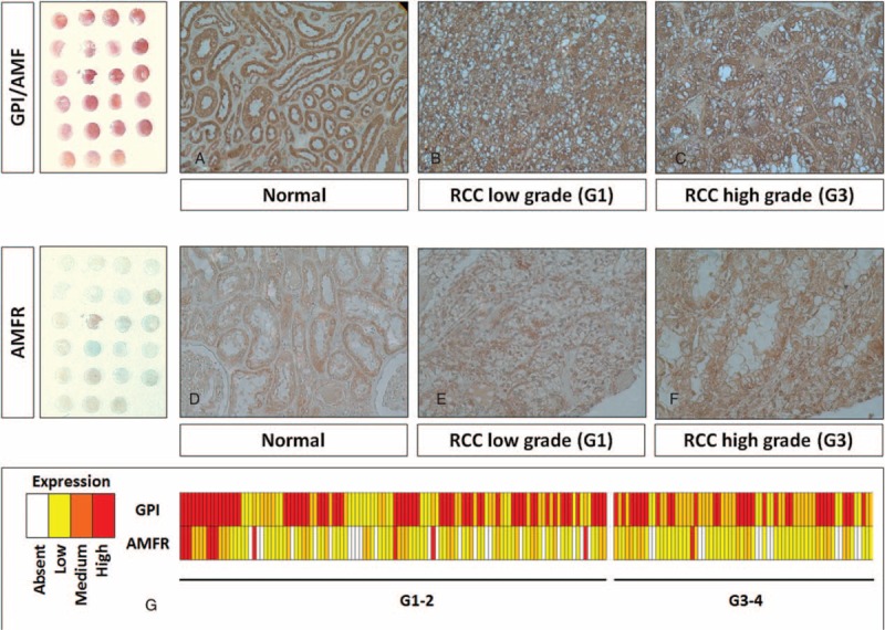
Immunohistochemical staining of GPI/AMF and AMFR proteins in tissue microarrays of human clear cell renal cell carcinoma (RCC) specimens. In normal kidney, GPI was predominantly localized in the cytoplasm of renal tubule cells, whereas it was absent in the glomeruli (Panel A). Clear cell RCC showed a stronger staining in cancer cells, with both a cytoplasmic and membranous pattern (Panels B and C). Similarly, AMFR expression was very low in normal kidney (Panel D), but showed higher levels in tumor tissue (Panels E and F). Heat map summarizing GPI and AMFR staining in 180 RCC patients (Panel G). Original magnifications 20×.
