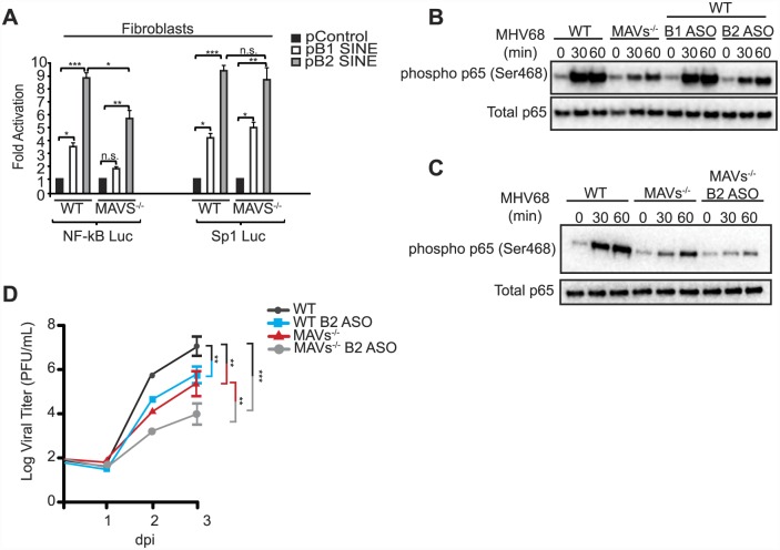
Fig 5. SINE RNAs signal through MAVS to activate NF-κB.
(A) Control, B1 SINE, or B2 SINE expression constructs were co-transfected with either the NF-κB or Sp1 reporter luciferase plasmid into WT and MAVS-/- fibroblasts. 48 h later luciferase levels were determined. (B) MAVS-/-, WT, and WT fibroblasts transfected with either B1 or B2 ASOs were infected with MHV68. At the indicated time points whole cell lysates were prepared and western blotted for phospho p65 (Ser468) and total p65. (C) WT, MAVS-/-, and MAVS-/- fibroblasts transfected with B2 ASO were infected with MHV68 and western blot analysis was performed at the indicated times for phospho p65 (Ser468) and total p65. (D) WT and MAVS-/- fibroblasts that are transfected with either control of B2 ASOs infected with MHV68 at an MOI of 0.05. Infection was allowed to progress for 72 h. Infectious virus produced was quantified by plaque assay in NIH3T3 cells.

