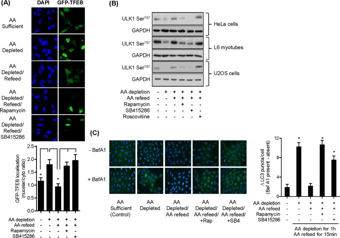
Figure 3. Effects of AA depletion/repletion and GSK3 inhibition upon markers of lysosomal biogenesis and autophagy.
(A) HeLa cells were cultured on coverslips and transfected with GFP-tagged TFEB. Twenty-four hours post transfection, cells were incubated in EBSS and EBSS+AA in the absence or presence of 100 nM rapamycin or 50 μM SB415286 as indicated and cells were fixed, imaged and analysed as described in the Materials and methods section. Asterisks indicate significant (P<0.05) changes between indicated bars. (B) HeLa cells, L6 myotubes and U2OS cells were subjected to incubation with EBSS containing or lacking AAs for 1 h or alternatively having been depleted of AAs incubated in EBSS containing a 1× AA mix (refeed) for 15 min in the absence/presence of 100 nM rapamycin, 50 μM SB415286 or 30 μM roscovitine as indicated. Cells were lysed and 30 μg of lysate protein was analysed by immunoblotting. (C) U20S cells were incubated as in (B) but in the absence or presence of 50 nM bafilomycin A1. Cells were fixed and nuclei (blue DAPI) and LC3 puncta (green fluorescence) visualized as described in the Materials and methods section. Asterisks indicate a significant difference between the indicated bars (A) or indicate a significant difference in LC3 puncta observed relative to the untreated AA-sufficient control (P<0.05).

