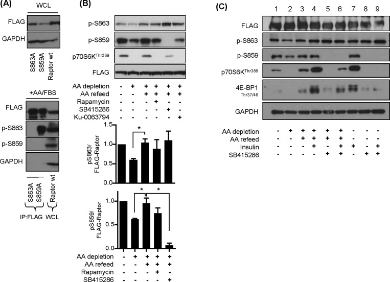
Figure 8. Effects of AA and insulin on raptor and p70S6K1 phosphorylation.
(A) HEK293T cells overexpressing FLAG–raptor, FLAG–raptor S859a or FLAG–raptor S863A mutant were lysed and FLAG-conjugated proteins were immunoprecipitated using anti-FLAG antibodies. Immunoprecipitates and 30 μg of lysate respectively were analysed by immunoblotting for the proteins indicated. (B) HEK293T cells overexpressing FLAG–raptor were incubated in EBSS containing or lacking AA for 1 h or alternatively having been depleted of AAs incubated in EBSS containing 1× AA mix (refeed) for 15 min. Cells were incubated with 100 nM rapamycin, 50 μM SB415286 or 1 μM Ku-0063794 for 15 min prior to and for 15 min during the AA-refeed period where indicated. Cells were harvested and 30 μg of protein was analysed by immunoblotting using the antibodies indicated. The lower panel represents quantification of Ser863 and Ser859 phosphorylation from at least three separate experiments (means ± S.E.M.) (C) HEK293 cells overexpressing FLAG–raptor were incubated in EBSS containing or lacking AA for 1 h ± 100 nM insulin (15 min) or alternatively having been depleted of AAs incubated in EBSS containing 1× AA mix (refeed) for 15 min ± 100 nM insulin. Cells were incubated with 50 μM SB415286 for 15 min prior to and for 15 min during the AA refeed period where indicated. Cells were harvested and 30 μg of protein was analysed by immunoblotting using the antibodies indicated.

