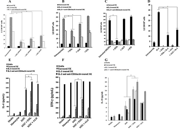
Figure 5. ZOL-treated osteoclasts and OSCSCs were resistant to NK-mediated cytotoxicity and secreted high levels of IFN-γ.

A. Highly purified NK cells (1 × 10⁶ cells/ml) were left untreated or treated with IL-2 (1000 units/ml) or a combination of IL-2 (1000 units/ml) and anti-CD16mAb (3 μg/ml) for 18 hours before they were added to 51Cr labeled osteoclasts (hOC) at various effector to target ratios. Osteoclasts were prepared as described in the Materials and Methods section and treated with ZOL (250 nM, 500 nM or 1 uM) for 30 minutes before they were used as target cells. NK cell mediated cytotoxicity was determined using a standard 4 hour 51Cr release assay. * The difference between IL-2 or IL-2+anti-CD16mAb stimulated NK cells treated with ZOL compared to IL-2 or IL-2+anti-CD16mAb stimulated NK cells without ZOL treatment is significant at P < 0.05 B. Osteoclasts were prepared as described in the Materials and Methods section and treated with ZOL, ALN or ETI (100 nM) for 30 minutes before used as target cells. NK cells were prepared as described in Fig. 5A and then added to 51Cr labeled osteoclasts at various effector to target ratios. NK cell mediated cytotoxicity was determined using a standard 4 hour 51Cr release assay. * The difference between IL-2 or IL-2+anti-CD16mAb stimulated NK cells treated with ZOL or ALN compared to IL-2 or IL-2+anti-CD16mAb stimulated NK cells without BP treatment is significant at P < 0.05 C. OSCSCs were treated with ZOL, ALN or ETI (1 μM) for 30 minutes before the addition of pre-treated NK cells, prepared as described in Fig. 5A. NK cell mediated cytotoxicity was determined using a standard 4 hour 51Cr release assay. * The difference between IL-2 stimulated NK cells treated with ZOL compared to IL-2 treated NK cells without BP treatment is significant at P < 0.05 D. Osteoclasts were differentiated from autologous monocytes as described in the Materials and Methods section for 17 days. NK cells were left untreated or treated with IL-2 (1000 units/ml) in the presence and absence of ZOL (500 nM) and ALN (500 nM) for 18 hours before they were added to 51Cr labeled osteoclasts at various effector to target ratios. NK cell mediated cytotoxicity was determined using a standard 4 hour 51Cr release assay. * The difference between IL-2 stimulated NK cells treated with ZOL or ALN compared to IL-2 stimulated NK cells without BP treatment is significant at P < 0.05. The lytic units 30/106 cells were determined using inverse number of NK cells required to lyse 30% of the osteoclasts or OSCSCs X100. E. Osteoclasts were treated as described in Fig. 5B and then added to untreated, IL-2 treated or IL-2+anti-CD16mAb treated NK cells at 1:3 ratio (NK: target cells). After an overnight incubation, the supernatants were harvested and the levels of IL-6 F. IFN-γ G. and IL-18 were measured with specific ELISAs. * The difference between IL-2 or IL-2+anti-CD16mAb stimulated NK cells treated with ZOL or ALN compared to IL-2 or IL-2+anti-CD16mAb stimulated NK cells without BP treatment is significant at P < 0.05 (E) * The difference between IL-2 stimulated NK cells treated with ZOL compared to IL-2 stimulated NK cells without BP treatment is significant at P < 0.05 (F) * The difference between untreated, IL-2 or IL-2+anti-CD16mAb stimulated NK cells treated with ZOL compared to those NK cells without BP treatment is significant at P < 0.05.
