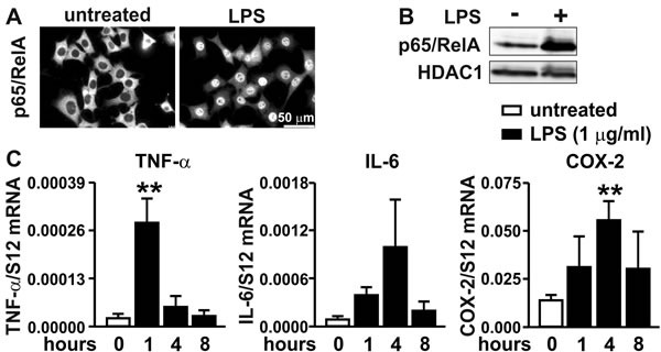
Figure 2. Inflammatory response of MC-38 cells.

A. Immunofluorescence microscopy of p65/RelA in MC-38 cells following treatment with 1 μg/ml LPS for 40 minutes. B. Immunoblot detection of p65/RelA and HDAC1 in nuclear extracts derived from MC-38 cells following stimulation with 1 μg/ml LPS for 1 hour. C. Kinetics of inflammatory induction (1 μg/ml LPS for the time periods indicated) of canonical NF-κB target genes quantified by RT-qPCR of TNF-α, IL-6 and COX-2 mRNA. Shown are mean values + SD of mRNA ratios relative to the constitutively expressed ribosomal protein S12 mRNA levels of n = 3 to 4 independent experiments; **, p < 0.01 (Student's t-test).
