
Figure 5. HIF signaling in p65/RelA knock-down cells.
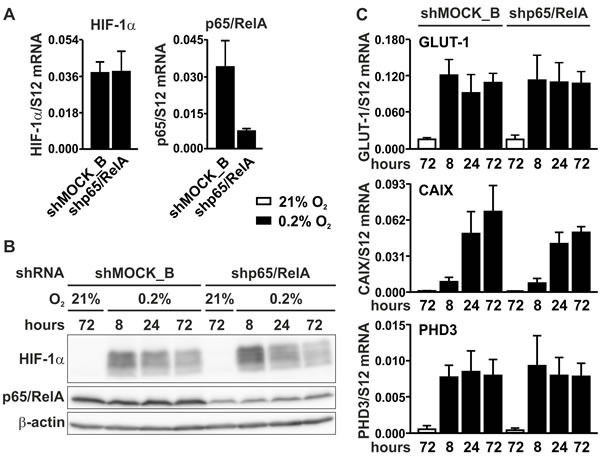
A. RT-qPCR analysis of HIF-1α and p65/RelA mRNA in MC-38 cells stably transfected with shp65/RelA or shMOCK_B negative control constructs. B. Immunoblotting of HIF-1α and p65/RelA protein after 8 to 72 hours of hypoxic exposure of shMOCK_B or shp65/RelA MC-38 cells. β-Actin served as loading control. C. RT-qPCR analysis of the canonical HIF target genes Glut1, Ca9 and Phd3 in shMOCK_B or shp65/RelA MC-38 cells cultured under normoxic or hypoxic conditions as indicated.