
Figure 6. p65/RelA signaling in HIF-1α knock-down cells.
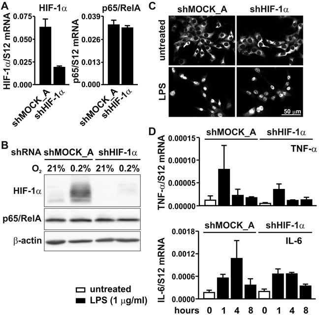
A. RT-qPCR analysis of HIF-1α and p65/RelA mRNA in MC-38 cells stably transfected with shHIF1α or shMOCK_A control constructs. B. Immunoblotting of HIF-1α and p65/RelA protein after 8 hours of hypoxic exposure of shMOCK_A or shHIF1α MC-38 cells. β-Actin served as loading control. C. Immunofluorescence microscopy of p65/RelA in shMOCK_A or shHIF-1α MC-38 cells treated with 1 μg/ml LPS for 40 minutes. D. RT-qPCR analysis of the canonical NF-κB target genes Tnfa and Il6 in shMOCK_A or shHIF-1α MC-38 cells upon treatment with LPS for 1 to 8 hours.