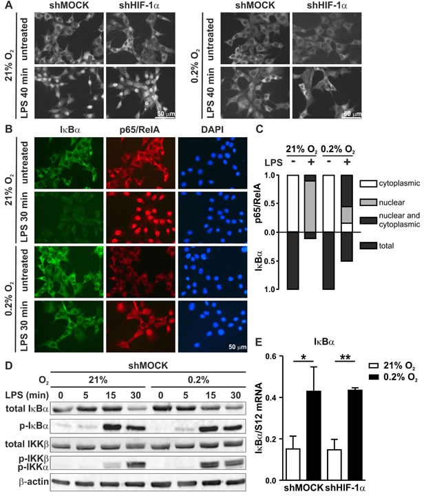
Figure 8. Inverse regulation of NF-κB and IκB under hypoxic conditions.

A. Immunofluorescence microscopy of p65/RelA in shMOCK and shHIF-1α MC-38 cells following exposure to 0.2% oxygen for 8 hours and/or 1 μg/ml LPS for the last 40 minutes before harvesting. B. Co-immunofluorescence microscopy of IκBα and p65/RelA in MC-38 cells (without GFP) exposed to 0.2% oxygen for 8 hours and/or 1 μg/ml LPS for the last 30 minutes before harvesting. C. For fractional quantification of the results shown in B., at least 200 cells of each experiment were classified according to the subcellular localization of p65/RelA and the expression of IκBα as indicated. D. Immunoblot analysis of total IκBα, phosphorylated IκBα, total IKKβ and phosphorylated IKKβ/α in MC-38GFP cells exposed to 0.2% oxygen for 8 hours and 1 μg/ml LPS for the last 5, 15 or 30 minutes as indicated. E. RT-qPCR analysis of IκBα mRNA levels in MC-38GFP cells exposed to 8 hours hypoxia and/or 1 hour LPS. Shown are mean values + SD of mRNA ratios relative to the constitutively expressed ribosomal protein S12 mRNA levels, normalized to the mean of each experiment (n = 3 independent experiments); *, p < 0.05; **, p < 0.01 (Student's t-test).
