Figure 1.
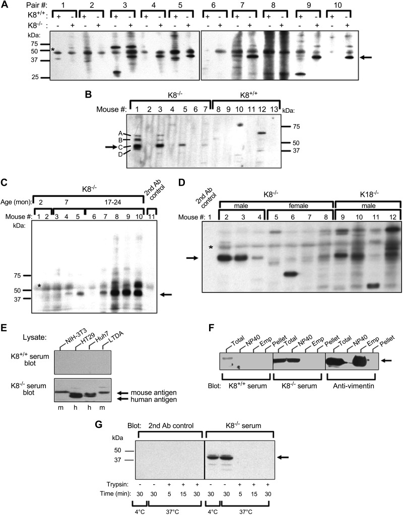
Autoantibodies are present in sera from aging K8−/− and K18−/− male mice. A–D) Sera from K8+/+, K8−/−, or K18−/− mice were analyzed by immunoblotting of total mouse liver lysates using a miniblotter setup. A) Ten age- and sex-matched K8+/+ and K8−/− mouse pairs (Pair #) were analyzed, and the arrow points to a protein band (antigen) that is consistently observed only in K8−/− sera. B) Only male mouse serum is analyzed to test for autoantibodies, and bands marked A–D highlight the 4 major autoantigens recognized using K8−/− but not K8+/+ sera. C) Sera from 10 male mice with the indicated ages were screened. D) Sera from aging (8 mo or older) K8−/− females and K18−/− male mice were tested for autoantibody presence using K8−/− as positive controls. The secondary (2nd) antibody control (lane 1) is indicated. C, D) Arrows highlight autoantigen C and asterisks (*), nonspecific signal. E) Mouse and human cell line lysates were tested by immunoblotting using sera from K8−/− and K8+/+ mice. F) NIH-3T3 fibroblasts were sequentially solubilized with NP-40 then Empigen and further analyzed (together with the total cell fraction or the post-Empigen pellet) by immunoblotting with K8+/+ or K8−/− serum, or anti-vimentin antibody as a control. G) NIH-3T3 NP-40 lysates were incubated with or without trypsin for the indicated times and then analyzed by immunoblotting using K8−/− serum or the secondary antibody control alone as a specificity control. Arrow highlights the autoantigen. Ab, antibody; Emp, Empigen; h, human; m, mouse; mon, month.

