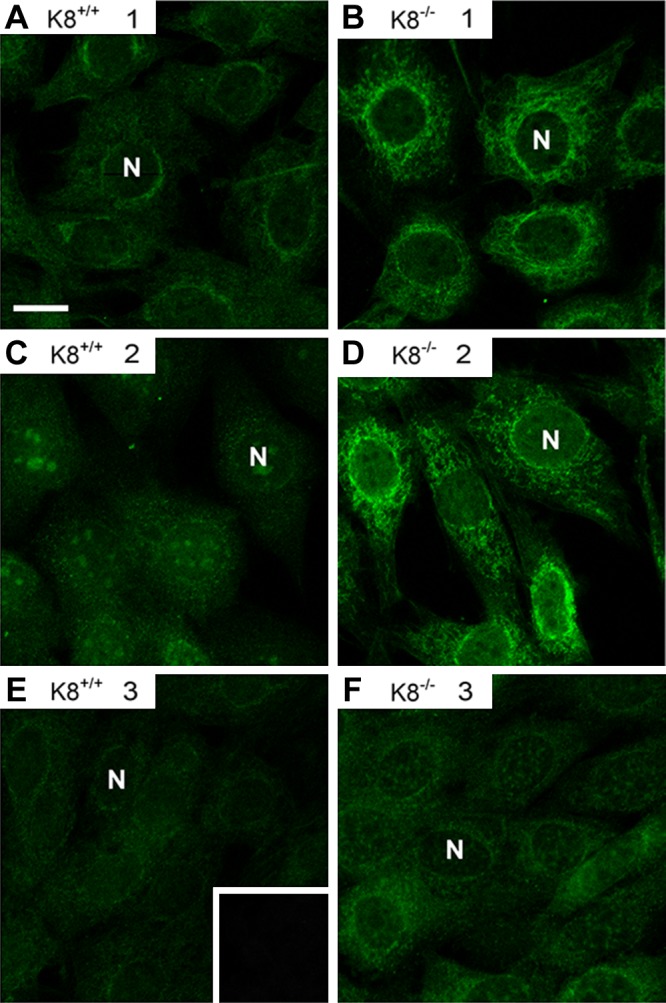
Figure 2.

K8−/− mouse autoantibodies manifest a cytoplasmic reticular staining pattern. K8+/+ (A, C, E) and K8−/− (B, D, F) sera from 3 mice (1–3) per genotype were used as primary antibodies to stain NIH-3T3 cells followed by FITC-labeled secondary anti-mouse antibody. The K8−/− sera were positive (B, D) or negative (F) for autoantibody reactivity as determined by immunoblotting. E) Insert represents secondary antibody alone control staining. N, nuclei.
