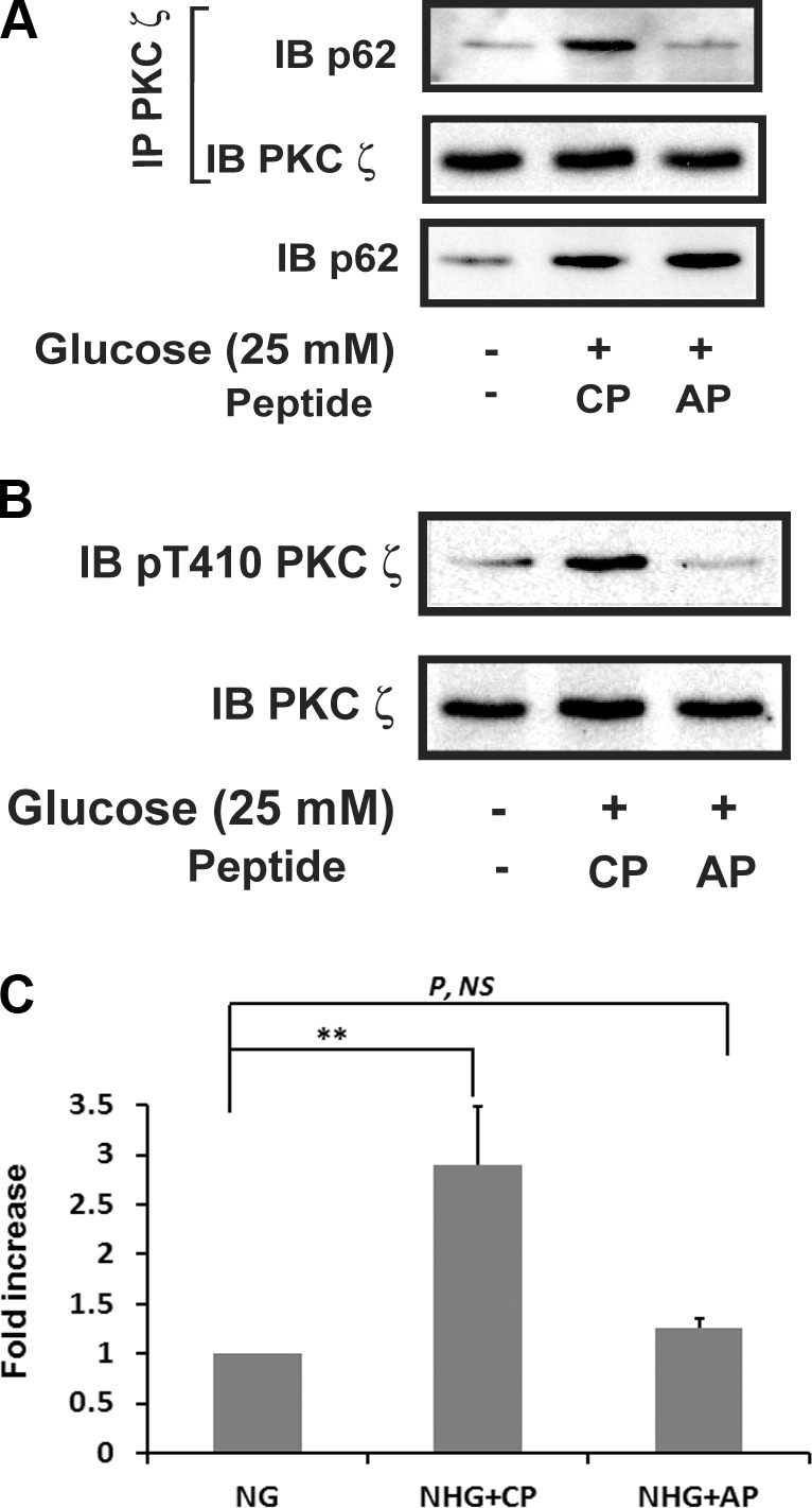
Figure 2.

Disruption of p62 and PKCζ association impairs hyperglycemia-stimulated PKCζ activation. VSMCs were cultured in DMEM containing normal glucose (5 mM) plus 10% FBS then serum deprived for 16 h before exposure to SFM with 25 mM glucose for 6 h in the presence of a control peptide (CP) or a disrupting peptide (AP) (10 μg/ml). A) Cell lysates were immunoprecipitated (IP) with an anti-PKCζ antibody and immunoblotted with an anti-p62 antibody. The blot was reprobed with an anti-PKCζ antibody. The same amount of lysate was immunoblotted with an anti-p62 antibody. B) Cell lysates were immunoblotted (IB) with an anti-pThr410 PKCζ antibody. To control for loading, the blots were stripped and reprobed with an anti-PKCζ antibody. C) PKCζ in vitro kinase activity was measured following the procedure described in Fig. 1C. P, NS, no significant difference in P value. **P < 0.01 indicates significant differences between 2 treatments.
