Figure 6.
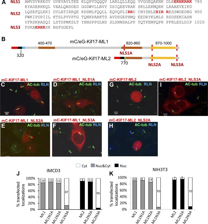
Identification of KIF17 NLS. A) C-terminal mouse KIF17 sequence showing 3 predicted NLS (NLS1–3, red). B) Schematic of NLS mutants of ML KIF17 peptides. Amino acids at positions 777–788 (NLS1A), 887–893 (NLS2A), and 1025–1028 (NLS3A) were replaced by alanines, with approximate locations in mC-KIF17-ML1 and mC-KIF17-ML2 peptides indicated (red vertical lines). C–F) Expression of mC-KIF17-ML1 and its 3 NLS mutants in IMCD3 cells. Primary cilia and basal bodies are labeled with anti–Ac-Tub (green) and anti-rootletin (blue) antibodies, respectively. Only mC-KIF17-ML1_NLS3A (F) was excluded from nuclei and localized in cytoplasm exclusively. Notably, mC-KIF17-ML1 and its 3 NLS mutants localized at distal tips of IMCD3 cell cilia. G–I) Expression of mC-KIF17ML2 and its 2 NLS mutants. Only mC-KIF17-ML2_NLS3A (I) was absent from nuclei and was instead distributed in cytoplasm. J and K) Statistical analyses of subcellular localizations of transfected KIF17ML1, KIF17ML2, and their NLS mutants expressed in IMCD3 (J) and NIH3T3 cells (K). Bars represent cytoplasmic (white), whole cell (gray), and nuclear (black) localizations, respectively.

