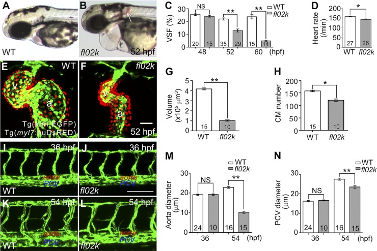
Figure 1.
Cardiovascular defects are evident in fl02k mutant embryos. A and B) Live images of a WT sibling and fl02k mutant embryo at 52 hpf. Arrow: interstitial hemorrhage in the mutant. C) VSF gradually declined from 48 to 60 hpf in fl02k embryos compared with their WT siblings. D) The heart rate was reduced in fl02k embryos at 52 hpf. E and F) Cardiac growth and expansion were compromised in fl02k mutants. The heart was stopped at end-diastole by 20 µM BDM treatment. Endocardial cells were labeled by Tg(kdrl:EGFP) (green), and myocardial nuclei by Tg(myl7:nuDsRED) (red). a, atrium; v, ventricle. G) Ventricular volume was reduced in fl02k mutants. The heart was stopped at end-diastole by 20 µM BDM. H) The number of CMs labeled by Tg(myl7:nuDsRED) decreased in fl02k mutants. I–N) The lumen of the aorta and PCV were formed at 36 hpf (J) but were not maintained at 54 hpf (L) in fl02k mutants compared with their WT siblings (I, K). I–L) Lateral views of trunk vessels of WT siblings and fl02k mutants in Tg(kdrl:EGFP) transgenic zebrafish. Lumens of the DA (L, M) and PCV (L, N) were narrower in fl02k embryos at 54 hpf, although the lumens were comparable in WT and fl02k mutant embryos at 36 hpf (I, J, M, N). The anterior is to the left in (I–L). The number of zebrafish embryos/group is shown in the histograms. *P < 0.05; **P < 0.01. Scale bars, 50 μm (E, F); 100 μm (I–L).

