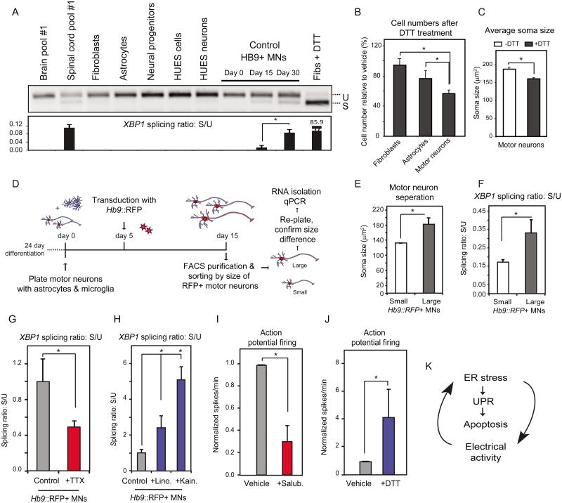
Figure 6. Human Motor Neurons Exhibit Increased Levels of Basal ER Stress That is Dependent on Their Physiological Activity.
(A) Purified Hb9∷GFP control MNs show higher levels of spliced XBP1 relative to other human cell types. Human spinal cord RNA also shows higher levels relative to brain RNA. U: unspliced, S: spliced. (B) Control MNs are more vulnerable to acute ER stress induction (DTT, 2mM) relative to fibroblasts or astrocytes, while (C) DTT treatment leads to a reduction in MN soma size (n=2, +/-SD, P<0.05). (D) Experimental strategy used to isolate MNs based on cell size. Control MN cultures were infected with the Hb9∷RFP virus on day 5. On day 15, RFP+ MNs were FACS-purified and sorted by size to separate small and large cells. (E) A subset of cells were re-plated to confirm soma size by measuring MAP2+ cell bodies (F). Basal levels of XBP1 splicing were assessed in RNA isolated from the remaining purified MNs showing that larger MNs had higher spliced XBP1 than smaller ones (n=3, +/-SEM, P<0.05). Treatment of MN cultures with (G) TTX reduces while (H) linopiridine and kainate increase XBP1 splicing respectively. Treatment with (I) salubrinal reduces, while (J) DTT increases action potential firing (n=3, +/-SEM, P<0.05). (K) ER stress, the UPR and electrical activity of MNs are connected.

