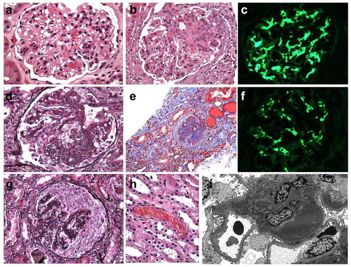
Figure 1. Pathologic features of IgA nephropathy by light microscopy, immunofluorescence and electron microscopy.
(a) The glomerulus has global mesangial proliferation with at least 4 cells per mesangial area. When >50% of glomeruli exhibit mesangial hypercellularity, the biopsy receives a score of M1 according to the Oxford/IgA MEST system (H&E, x600).
(b) Segmental endocapillary proliferation obliterates capillary lumina (score E1 when a biopsy contains one or more such lesions). The adjacent glomerular segments have mild mesangial hypercellularity (H&E, x600).
(c) The stain for IgA is intense and globally outlines the mesangial framework of the glomerulus (immunofluorecence, x600).
(d) Segmental glomerular scarring develops as postinflammatory sclerosis, mimicking the changes in focal segmental glomerulosclerosis. (score S1 when a biopsy contains one or more such lesions), (Jones methenamine silver, x600).
(e) A case with high chronicity contains globally sclerotic glomeruli and exhibits more than 50% tubular atrophy/interstitial fibrosis (score T2), (Masson trichrome, x200).
(f) The immunofluorescence staining for C3 is similar in distribution as the mesangial staining for IgA (shown from the same glomerulus as in 1C) but exhibits weaker intensity and a more punctate, granular texture (immunofluorescence, x600).
(g) A severe example has a cellular crescent that compresses the glomerular tuft. Global mesangial expansion is present (Jones methenamine silver, x400).
(h) One or more red blood cell casts are commonly encountered at biopsy and may be numerous, especially in cases with gross hematuria and acute tubular injury (H&E, x600).
(i) By electron microscopy, large mesangial deposits elevate the glomerular basement membrane reflection over the mesangium, bulging towards the urinary space. This deposit involves the entire mesangium but is most prominent in the paramesangial region, beneath the GBM reflection. The mesangial cellularity is increased but the capillary lumen is patent (electron micrograph, x5000).

