Fig. S1.
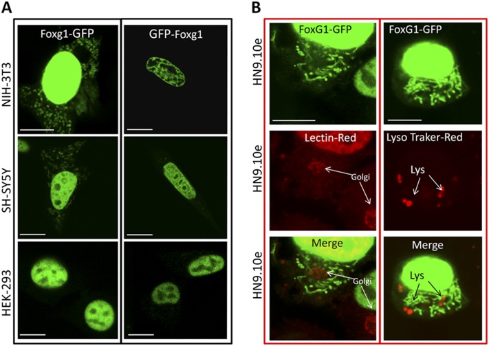
Subcellular localization of transiently transfected Foxg1-GFP and GFP-Foxg1 in different cell lines. The pictures (A) indicate that only Foxg1-GFP localizes both in the nucleus and at mitochondria. It is worth noting that the GFP N-terminal fusion can inhibit the correct mitochondrial localization. The only exception was found in HEK293 cell line, where Foxg1-GFP does not present any mitochondrial localization, but only a nuclear localization. (Scale bar, 5 µm.) (B) Foxg1-GFP (green) does not overlap with lysosomes labeled with lysotracker (Red DND-99 molecular probe), nor with Golgi labeled with lectin (red).

