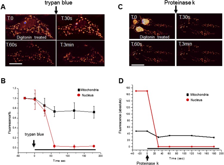
Fig. S6.
Submitochondrial localization of Foxg1-GFP in transfected living cells. (A) Representative time-lapse of a Foxg1-GFP overexpressing living NIH 3T3 cell permeabilized with digitonin and treated with trypan blue. (Scale bar, 5 µm.) These experiments are resumed in B (mean ± SE, n = 5 in three independent experiments). (C) Time-lapse of NIH 3T3 cells permeabilized with digitonin and treated with proteinase K. (D) Exemplificative time course of mitochondrial and nuclear GFP fluorescence in a permeabilized cell imaged in C.

