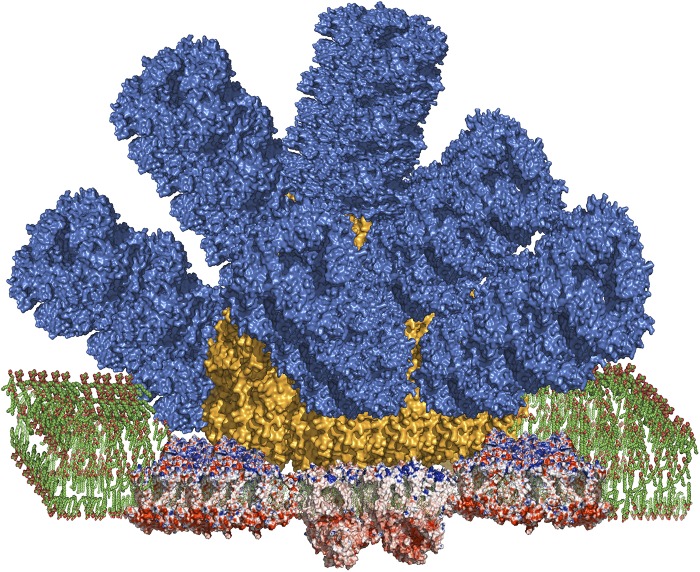
Fig. 1.
Structural model of a phycobilisome antenna complex associated with both photosystem I and photosystem II in a megacomplex. The model is based on X-ray crystal structures of the phycocyanin and allophycocyanin proteins and the reaction centers. The relative positions and orientations of the phycobilisome and reaction centers are based on chemical cross-linking and MS studies (11). Color coding is as follows: phycocyanin, blue; allophycocyanin, orange; reaction centers are shown in red (negative charged residues), blue (positively charged residues), and tan (uncharged residues). Photosystem II is present as a dimeric complex directly under the core of the phycobilisome, with the downward-pointing lobes that make up the accessory subunits of the oxygen-evolving system. Photosystem I is present as two copies of a trimeric complex that flanks the central attachment site of the phycobilisome on photosystem II. The lipids of the thylakoid membrane bilayer are shown in green. Image courtesy of Haijun Liu (Washington University in St. Louis, St. Louis).

