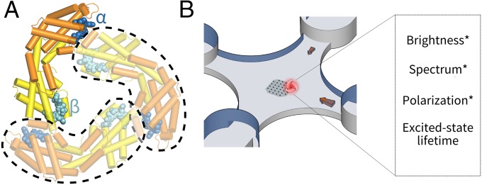
Fig. 1.
Experimental scheme. (A) Crystal structure of the model antenna protein used in this study: APC (PDB ID code 1ALL) is a trimer with C3 symmetry. Each constituent monomer (circled in dashed lines) binds two heterogeneous PCB pigments (α, blue; β, cyan). In the trimer, a β-pigment is brought to close proximity to the α-pigment of a neighboring monomer. (B) A single-molecule fluorescence assay to probe the photodynamics of individual APCs in solution. Molecules are held in an anti-Brownian ELectrokinetic trap and interrogated by polarization-resolved multiparameter fluorescence spectroscopy.

