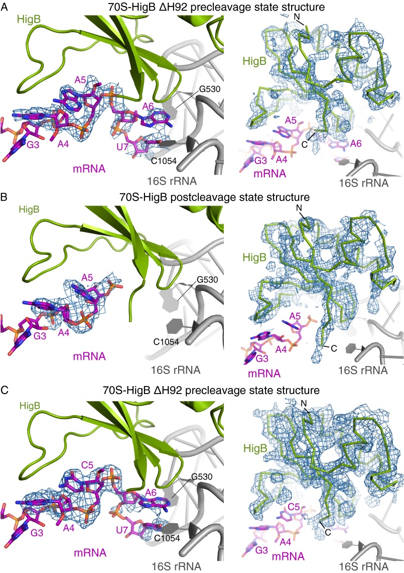
Fig. S1.
Quality of 70S-HigB bound maps and models. Unbiased Fo-Fc difference electron density (contoured to 2σ unless noted otherwise) for both the mRNA (Left) and HigB (Right) for 70S ribosome structures bound to HigB. (A) A 3.4-Å X-ray crystal structure of the 70S-HigB ΔH92 precleavage state containing an A-site AmAmAm lysine codon. (B) A 3.3-Å X-ray crystal structure of the 70S-HigB postcleavage state containing an A-site AAA lysine codon. (C) A 3.1-Å X-ray crystal structure of the 70S-HigB ΔH92 precleavage state containing an A-site AmCmAm lysine codon. Here, unbiased Fo-Fc difference electron density is contoured to 1.5σ and 2σ the mRNA (Left) and HigB (Right), respectively.

