Fig. S2.
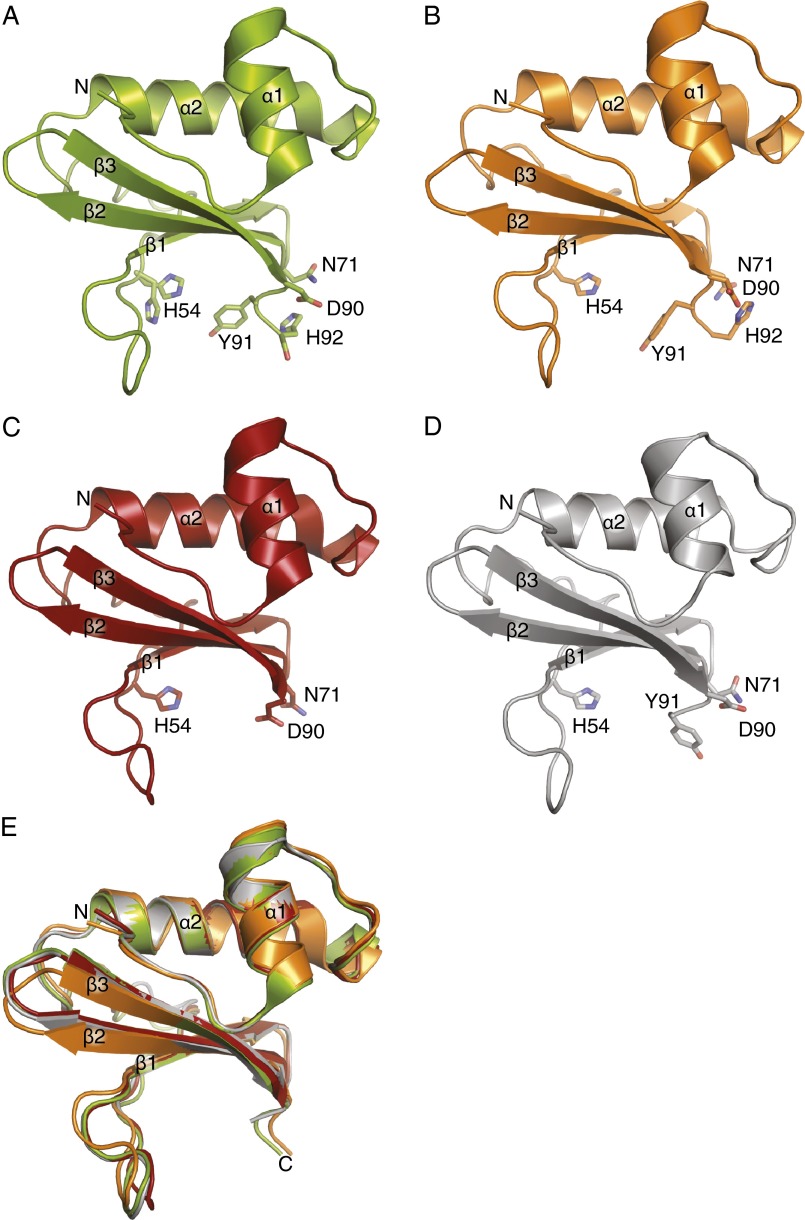
Comparison of HigB in different states. (A) A 1.25-Å X-ray crystal structure of free HigB with active site residues shown as sticks. (B) A 2.8-Å X-ray crystal structure of the HigBA complex was solved but only HigB is depicted (PDB code 4MCT). (C) Structure of HigB from the 2.1-Å X-ray crystal structure of the HigBA complex (PDB code 4MCX). (D) A 3.4-Å X-ray crystal structure of HigB ΔH92 bound to the 70S ribosome in the precleavage state. (E) Overlay of all four HigB structures demonstrating their overall similar fold (rmsd 0.4–1.0 Å for residues 1–92).

