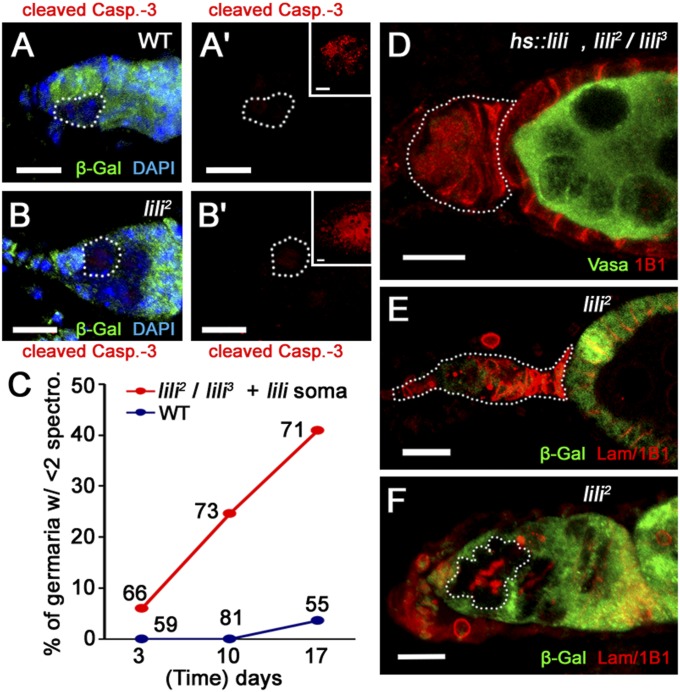
Fig. 2.
Loss of lili mutant GSCs is not caused by cell death or cell competition. (A and B) Germaria containing either WT control (A) or lili mutant (B) clonal GSCs (β-gal negative; dashed lines) do not show any activated Caspase-3 staining (red) (A′ and B′). Positive cell death control (anti-activated Caspase-3 Ab; red) stage 7–8 egg chambers are shown in Insets (Fig. S4). Clone tissue is marked by absence of β-gal (green). (C) GSCs are lost over time in hs-Gal4/UASt-liliA; lili2/lili3 females. (D) Example of a “GSC-depleted” germarium (dashed outline) stained for the germ line (Vasa; green) and membranes/spectrosomes/fusomes (1B1; red) from a 10-d-old hs-Gal4/UASt-liliA; lili2/lili3 female. (E) A similarly depleted germarium (dashed outline) attached to a lili2 mutant clonal egg chamber (β-gal present in the soma but not the germ line) stained for the spectrosome, fusome, and cell membranes marker combination Lamin C/1B1 (red). (F) Example of a GSC-depleted germarium showing developing lili2 clonal cysts (dashed outline; β-gal absent) at the site normally occupied by GSCs, staining is with anti-LamC/anti-1B1 combination (red; branched fusome is clearly visible in clonal cyst). (Scale bars: 10 μm.)

